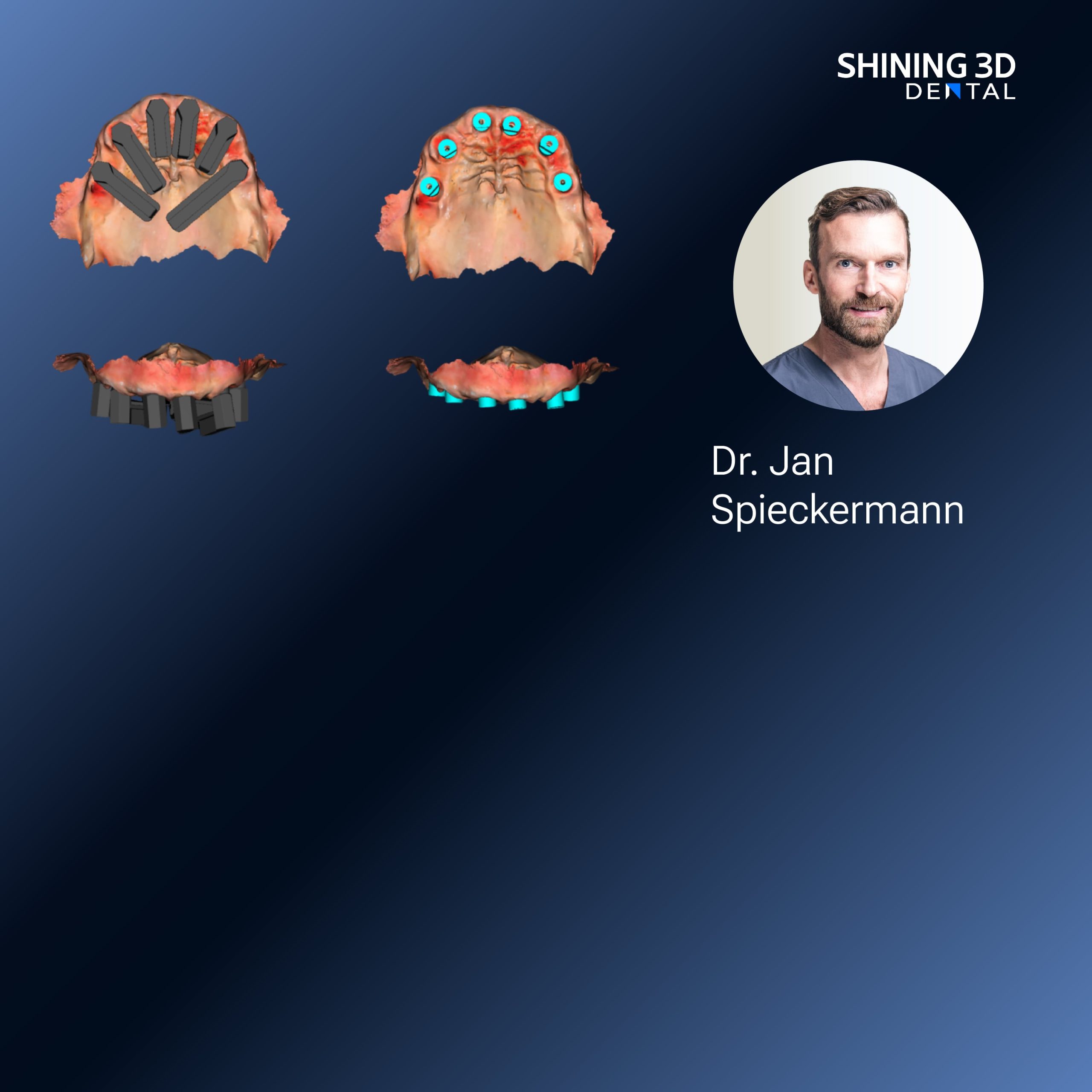
Introduction — data framing and scope
This note presents measured questions and practical answers about digital denture workflow, shaped by data and laboratory practice rather than marketing assertions. Early decisions in scanning and registration define fit more than later polish. For example, reliable capture of occlusion and vertical dimension hinges on a correct jaw relation record, and modern workflows often integrate dedicated capture devices to reduce manual adjustment. Recent demonstrations at IDS Cologne emphasized interoperability and clinical validation as decisive factors; that event remains a useful reference for what vendors show and what clinicians demand. My observations derive from prolonged collaboration with prosthodontic clinics and digital laboratories: familiarity with scanner output files, lab milling parameters and print post-processing informs the recommendations below.

Scanning — what to measure and why it matters
Scan quality is not binary. Measure three characteristics: surface resolution, repeatability, and true-to-geometry capture of soft tissue margins. Handheld intraoral scanners can deliver adequate surface detail for denture bases if soft tissue motion is managed; laboratory bench scanners give superior repeatability when working from impressions or casts. Expect variation in soft-tissue areas; use controlled lighting and minimal patient movement to lower that variation. Acceptable practice: verify scans against a physical reference or a stone cast when possible, and preserve original scan files for traceability.
Design — parameters that change outcomes
Design choices must be explicit and documented: base thickness, relief at frenula and vestibules, posterior palatal seal placement, and occlusal schemes. Software algorithms may propose automated tooth set-ups; treat these as starting points. Critical control points require human verification — primarily the occlusal vertical dimension and centric relation. Export and review occlusion in a neutral file format. Where software supports it, annotate design intent inside the CAD file so the technician and clinician share the same expectations.
Production — choosing between additive and subtractive
Both modalities produce clinically acceptable dentures when process control is strict. Milling offers dense polymethyl methacrylate (PMMA) blocks with predictable mechanical properties; additive manufacturing allows complex undercuts and internal retention features. Control variables: layer orientation for prints, post-curing protocol, milling bur condition, and material batch traceability. Document the chosen parameters for each case. Do not assume identical fit across technologies; validate fit with a trial insertion or try-in when possible.
Verification and quality metrics — what to check before delivery
Implement a short checklist: marginal adaptation to soft tissue, occlusal contacts, flange extensions, and esthetic positioning of teeth. Use objective measures when available: digital superimposition to quantify deviation in the tens of micrometers, pressure-indicating paste to map contact distribution, and photographic records for esthetics. Record patient feedback at the first insertion and after two weeks; those data points predict final adjustment workload.
Alternatives and comparative evaluation
Conventional workflows remain viable: functional impressions and laboratory processing provide excellent tissue adaptation for many patients. Hybrid workflows — intraoral scanning for bite and try-in, analog impressions for tissue capture — can reduce rework while preserving clinical control. Evaluate alternatives by their effect on chair time, number of adjustments, and reproducibility across cases rather than by feature lists alone.
Common pitfalls and how to prevent them
Most failures follow predictable patterns: poor soft-tissue capture, unverified occlusal registration, and overlooked post-processing variability. Prevent these by standardizing capture protocols, requiring a verified jaw relation record when indicated, and logging all production parameters. Train at least two staff members to handle scanning and design to avoid single-point technique dependency. Keep a short corrective-action log for each returned case to spot systemic issues.
Final synthesis
Data-driven practice demands measurement at each stage: quantify scan fidelity, document design decisions, control production parameters, and track patient outcomes. These steps reduce adjustments and make results predictable. The practical value appears when a laboratory or clinic moves from anecdote to reproducible process; companies that show validated solutions and standard protocols at events like IDS Cologne help set benchmarks. When a workflow yields consistent fit and minimal remakes, it becomes a tool rather than a novelty, as demonstrated by adoption in regional specialty centers. That practical consistency is precisely the outcome you seek from a capable partner such as SHINING 3D DENTAL, whose product and process documentation align with the measurable controls described above.